| 2005 |

|
YEAR BOOK |
Cork Institute of Technology
|
Acoustic signature analysis of drills used in critical surgical procedures
|
(Figure 1)
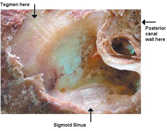
The temporal bone, situated around the ear, includes the mastoid region which is a mixture of cellular and solid bone. The mastoid frequently requires surgical dissection to eliminate diseases or infections or to provide access to other organs. The dissection process uses specialised high-speed drills. As the drill penetrates inwards, the surgeon uses anatomical knowledge and changes in tissue colour and drill pitch to avoid accidental damage.
Recently, a medical�electronic partnership was formed between CIT and SIVH to explore the possibility of using acoustic analysis to enhance the reliability and safety of the process. A successful application was made to Science Foundation Ireland (SFI) which resulted in funding for a one-year pilot project. This work will make preliminary investigations into the application of acoustic signature analysis and modelling theory for use in tracking drill depth. Some work will also be done on the integration of drill position information into a virtual environment which incorporates patient-specific scan information, such as Computer Aided Tomography (CAT) and Magnetic Resonance Imaging (MRI). This would provide a visual surgical aid for preoperative or live surgery. As a training mechanism, it would also assist portability of virtual skills to theatre skills.
The skill used in temporal bone dissection is achieved through cadaveric bone specimens in the first instance followed by live experience in the operative environment. Cadaveric dissection introduces the trainee surgeon to anatomical variations, the spatial relationships between adjacent structures and use of the operating drill. More recently, virtual reality environments have been developed which show on-screen drill progression. The trainee holds a drill-type transducer which simulates an actual drill. Background, computer generated, synthesised drill noise helps create a real acoustic environment. With a lack of availability of modelling work on the drill acoustic signature, the latter usually fails to be realistic thus diminishing the fidelity of the simulation.
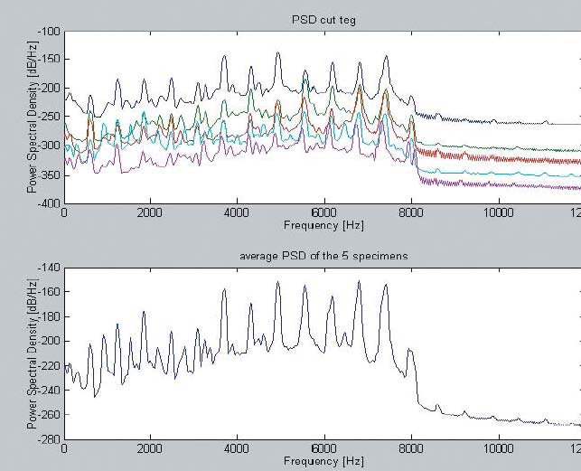
Typically, the drilling process will first drill through the thick bone at the mastoid surface and then proceed to the bony covering of the tegmen tympani (part of the ear and linked to the brain). It is drilled to wafer-thin thickness. Then, the bony covering of the sigmoid sinus (a system of veins which drains blood from the brain) is also drilled to wafer-thin thickness. Identification of the tegmen tympani and the sigmoid sinus is largely achieved using visual cues. Specific to the tegmen, progressive thinning of the bone is accompanied by a subtle acquisition of a pink hue and an acoustic change in the drill pitch. A similar change occurs when the bone overlying the sigmoid sinus becomes thin. The sound changes provide a timely warning to the surgeon of the proximity to these underlying structures. No study has attempted to objectively characterize this change in sound. With the continued depletion of available cadaveric temporal bones for dissection training, there is a real need to generate acoustic models for training purposes.
In the last ten years, robotic systems for surgical practices and training simulations have received much attention. Orthopaedic procedures, in particular hip and knee replacements and spinal fusion, were the first target areas for the development of robot applications. Compared with soft tissues, bones are relatively easy to manipulate and deform little during cutting. In relation to training and practice of temporal bone dissection, the solution is via a human-automation partnership. The challenge to the electro-medical team is to create accurate acoustic models corresponding to variations in bone and tissue types, drill types and drill speeds. These models can then be used to optimise the speed of the operation without compromising critical safety limits. They can also be used to create acoustically accurate virtual training environments and, in conjunction with patient-specific data from CAT, MRI and 2D scans, to create live, flexible and detailed screen images of the surgical target area.
(Figure 2)
Contact: Dr. Joseph Connell,
Advanced Control Group, Department of Electronic Engineering,
Cork Institute of Technology;
E-mail: [email protected] ; Web: www.cit.ie